Basal Cell Carcinoma Pictures: Recognize Early Warning Signs
When a spot on your skin just won’t heal, it’s easy to brush it off as a stubborn pimple or a scratch that’s taking its time. But for millions each year, that persistent bump turns out to be basal cell carcinoma (BCC)—the most common skin cancer in the United States, with over 3.6 million cases diagnosed annually.
BCC is the most common skin cancer in the U.S.: Over 3.6 million cases per year ·
BCC rarely spreads to other organs: Less than 1% of cases ·
Most BCCs develop on sun-exposed areas: Face, ears, scalp, neck, arms, legs ·
BCC can be cured if treated early: 95-99% cure rate
Quick snapshot
- BCC is the most common skin cancer, making up about 80% of non-melanoma cases (Skin Cancer Foundation)
- Most BCCs appear on sun-exposed areas, especially the head and neck (Mayo Clinic)
- Early detection leads to a cure rate >99% (Skin Cancer Foundation)
- Exact time from BCC onset to metastasis is unpredictable if it occurs (Mayo Clinic)
- Individual growth rate varies widely and is not well understood (Mayo Clinic)
- Specific triggers for aggressive BCC subtypes remain unknown (Skin Cancer Foundation)
- 0-3 months: Small pearly bump or non-healing sore appears
- 6-12 months: Lesion may enlarge, develop central ulceration or crusting
- 1-2 years: Moderate local invasion; possible pain or bleeding
- 5+ years: Advanced local invasion; potential bone or cartilage involvement
- See a dermatologist for any spot that does not heal within 4 weeks (American Academy of Dermatology)
- A biopsy can confirm the diagnosis (Mayo Clinic)
- Treatments include surgical excision, Mohs surgery, cryotherapy, and topical creams (American Academy of Dermatology)
The table below outlines key facts about basal cell carcinoma, including its prevalence, metastasis rate, and cure potential.
| Factor | Value |
|---|---|
| Most common skin cancer type | Yes, 80% of all non-melanoma skin cancers (Skin Cancer Foundation) |
| Annual cases in U.S. | Approximately 3.6 million (Skin Cancer Foundation) |
| Metastasis rate | Less than 0.5% (Mayo Clinic) |
| 5-year cure rate with early treatment | >99% (Skin Cancer Foundation) |
| Common body sites | Head, neck, face (80%), trunk (15%), extremities (5%) (Mayo Clinic) |
| Typical growth period before treatment | Months to years (median 2-3 years) (Healthgrades) |
What does stage 1 basal cell carcinoma look like?
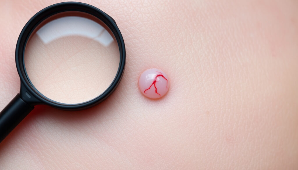
Stage 1 BCC is the earliest invasive stage, and catching it here makes all the difference. The tumor is small — usually less than 2 cm — and often appears as a pearly or translucent bump that may have tiny visible blood vessels (telangiectasias). On lighter skin it looks pink or red, while on darker skin it can be brown or glossy black (Mayo Clinic). Many patients mistake it for a pimple that never heals (AIM at Skin Cancer Foundation).
Characteristics of early BCC appearance: pearly, translucent bump
- Small, often less than 2 cm (Skin Cancer Foundation)
- Shiny, skin-colored translucent bump (Mayo Clinic)
- May have visible blood vessels (telangiectasias) (Skin Cancer Foundation)
The implication: Stage 1 BCC often hides in plain sight. Because it looks like a harmless blemish, many people wait months before seeking a dermatologist — and that delay matters.
Common vs atypical presentations on different body sites
- On the face: often a pearly nodule on the nose, eyelids, or cheeks (Mayo Clinic)
- On the trunk or arms: may appear as a reddish patch or flat scaly area (American Academy of Dermatology)
- On dark skin: pigmented BCC can be mistaken for a mole (Mayo Clinic)
Color variations: flesh-toned, pink, red, or dark pigmented
- Flesh-toned or pearly white on fair skin (Skin Cancer Foundation)
- Pink or red on sun-damaged skin (Skin Cancer Foundation)
- Brown, black, or blue in people with darker skin (Mayo Clinic)
The pattern: Recognizing these subtle differences across skin tones and body sites is critical for early detection. The earlier a BCC is identified, the simpler the treatment and the better the cosmetic outcome.
What are the five warning signs of basal cell carcinoma?
These five signs, established by the Skin Cancer Foundation, help you differentiate BCC from harmless spots. Remember: BCC does not follow the ABCDE rule of melanoma — its warning signs are different.
- Open sore that bleeds, oozes, or crusts and does not heal for weeks (Skin Cancer Foundation)
- Reddish patch or irritated area on the face, chest, back, or arms that may crust or itch (Skin Cancer Foundation)
- Shiny bump or nodule that is pearly, pink, red, or white (Skin Cancer Foundation)
- Pink growth with a rolled, elevated border and a crusted indentation in the center (Skin Cancer Foundation)
- Scar-like area that is white, yellow, or waxy, with poorly defined edges (Skin Cancer Foundation)
What this means: These signs should not be ignored. The American Academy of Dermatology recommends checking your skin monthly and seeing a dermatologist for any spot that is new, changing, or not healing within 4 weeks.
Where is the most common place to get basal cell carcinoma?
BCC overwhelmingly chooses the head and neck — up to 80% of cases occur there (Mayo Clinic). The nose is the single most common site (Skin Cancer Foundation).
- Head and neck: face, ears, scalp, lips (Mayo Clinic)
- Trunk: shoulders, back (about 15%)
- Extremities: arms, legs (about 5%)
- On darker skin: BCC can appear on less typical areas like the trunk and legs (Mayo Clinic)
The pattern: Sun exposure drives these numbers. The face and neck receive the most UV damage over a lifetime, making them ground zero for BCC.
When to worry about basal cell carcinoma?
Most BCCs are slow-growing and rarely life-threatening, but certain red flags call for immediate attention. Worry signs include rapid growth, bleeding, pain, or numbness (Skin Cancer Foundation).
- Any lesion that grows noticeably over a few weeks (Mayo Clinic)
- Spontaneous bleeding or crusting that recurs (Skin Cancer Foundation)
- Altered sensation (itch, tenderness, numbness) around the spot
- Size larger than 2 cm or duration longer than 1-2 years (Healthgrades)
The catch: BCC becomes dangerous when it invades deeper structures — nerves, bone, cartilage. That is rare but can lead to disfigurement and complex surgery.
How long does basal cell carcinoma take to spread?
BCC is notoriously slow. The typical growth timeline spans months to years, with a median of 2-3 years before treatment (Healthgrades). Metastasis — spread to lymph nodes or distant organs — occurs in less than 0.5% of cases (Mayo Clinic).
That said, certain aggressive subtypes (morpheaform, basosquamous) can grow and spread faster. The Skin Cancer Foundation notes that neglected tumors can eventually invade bone and cartilage.
The very low metastasis rate is reassuring, but local destruction is the real risk. A BCC left untreated for years can eat through skin, muscle, and bone, requiring extensive reconstruction.
The implication: Even though metastasis is rare, the potential for local damage means that any non-healing spot should be evaluated promptly to avoid complex surgery down the line.
Basal cell carcinoma stages pictures
BCC is staged by size, depth, and spread. The TNM classification system is standard (Skin Cancer Foundation). Here is a visual guide to each stage:
Stage 0 (Tis): Carcinoma in situ
- Abnormal cells only in the epidermis; no invasion
- Often appears as a flat, scaly, red or brown patch
Stage 1 (T1): Small, less than 2 cm
- Pearly bump or nodule, often mistaken for a pimple (Mayo Clinic)
- May have visible blood vessels
Stage 2 (T2): Larger than 2 cm or deeper invasion
- Often ulcerated or crusted (Skin Cancer Foundation)
- More obvious vascular pattern
Stage 3 (T3): Advanced local invasion
- Tumor invades deep dermis, subcutaneous tissue, possibly bone or cartilage
- Pain, bleeding, numbness common
- Requires extensive surgery or radiation (Mayo Clinic)
Stage 4 (T4): Metastatic BCC
- Spread to lymph nodes or distant organs
- Extremely rare (<0.5% of cases) (Mayo Clinic)
- Systemic symptoms: weight loss, fatigue
- Targeted therapy options available (Skin Cancer Foundation)
The implication: Each stage shows a clear progression in size, depth, and symptoms. Stage 1 is easily cured; stage 4 is rare but serious.
How long is too long to leave a basal cell carcinoma?
Dermatologists agree: any spot that persists longer than 4 weeks without healing warrants a professional look (American Academy of Dermatology). The longer you wait, the deeper the invasion and the more complex the treatment.
- After 3 months: lesion may enlarge and ulcerate
- After 1 year: possible deep dermal invasion
- After 2 years: can involve cartilage or bone (Healthgrades)
Delaying treatment increases the chance of a larger surgical excision, possible nerve damage, and a less cosmetic outcome. For a stage 1 BCC caught early, a simple 15-minute excision can cure it permanently.
The catch: Time is a critical factor in BCC management. The longer a lesion remains untreated, the higher the likelihood of requiring more invasive procedures and experiencing functional or aesthetic consequences.
Six skin cancer types, one key distinction: BCC rarely kills, but it can disfigure. Melanoma kills more often but is less common. SCC sits in between. The table below shows the critical differences you need to know.
| Feature | Basal Cell Carcinoma | Squamous Cell Carcinoma | Melanoma |
|---|---|---|---|
| Appearance | Pearly or pigmented bump, often with telangiectasias (Skin Cancer Foundation) | Firm, red, scaly or crusted patch (American Academy of Dermatology) | Irregular, asymmetrical, multicolored mole or spot (Skin Cancer Foundation) |
| Common sites | Head, neck, face (80%) (Mayo Clinic) | Sun-exposed areas: ears, lips, hands (American Academy of Dermatology) | Anywhere on body; more common on back in men, legs in women (Skin Cancer Foundation) |
| Growth rate | Slow (months to years) (Healthgrades) | Moderate (weeks to months) (American Academy of Dermatology) | Can be rapid (weeks) (Skin Cancer Foundation) |
| Metastasis risk | <0.5% (Mayo Clinic) | 1-10% (American Academy of Dermatology) | High if not caught early (Skin Cancer Foundation) |
| Treatment | Surgery, Mohs, cryotherapy, topical (American Academy of Dermatology) | Surgery, radiation (if high risk) (American Academy of Dermatology) | Surgery, immunotherapy, targeted therapy (Skin Cancer Foundation) |
Upsides and downsides of treating BCC early vs delaying
Upsides
- Cure rate >99% with early treatment (Skin Cancer Foundation)
- Simple outpatient excision leaves minimal scarring
- No risk of metastasis if caught at stage 1 (Mayo Clinic)
- Lower cost and faster recovery
Downsides
- Delayed diagnosis can lead to local invasion and disfigurement (Healthgrades)
- Advanced BCC may require Mohs surgery or radiation
- Long-term neglect can result in nerve damage or bone involvement (Skin Cancer Foundation)
- Larger tumors leave more noticeable scars
The pattern: The decision to treat early is clear-cut — the benefits far outweigh the risks. Delaying only increases the complexity and potential for harm.
How to examine your skin for BCC: a step-by-step guide
The American Academy of Dermatology recommends a monthly self-exam. Here is a simple routine:
- Use a full-length mirror and a hand-held mirror for hard-to-see spots
- Check your face, ears, scalp (part your hair), neck, and lips (Mayo Clinic)
- Examine arms, hands, underarms, chest, abdomen, and back
- Check legs, feet (including soles and between toes)
- Look for any new, changing, or non-healing spot — especially pearly bumps or sores that crust (Skin Cancer Foundation)
- If you find something suspicious, take a photo and schedule a dermatology appointment within 2 weeks
The catch: Self-exams can miss early BCC, especially on the scalp and back. Annual professional skin checks are recommended for anyone over 50 or with a history of sunburns (American Academy of Dermatology).
What we know vs what’s still unclear about BCC
Confirmed facts
- BCC is the most common form of skin cancer (Skin Cancer Foundation)
- Sun exposure and UV radiation are primary risk factors (Mayo Clinic)
- Most BCCs appear on sun-exposed areas (Mayo Clinic)
- Early-stage BCC is highly curable (Skin Cancer Foundation)
- Metastasis is very rare (<0.5%) (Mayo Clinic)
What’s unclear
- Exact time from BCC onset to metastasis (if ever) is unpredictable
- Individual growth rate varies widely
- Specific triggers for aggressive subtypes are not fully understood
- Whether sunscreen alone can prevent all BCCs remains debated (American Academy of Dermatology)
The pattern: While many aspects of BCC are well understood, the variability in growth and behavior means that individual cases can still surprise clinicians. This underscores the need for regular monitoring and prompt evaluation of any suspicious lesion.
Expert perspectives on BCC pictures
“Early BCC often looks like a pearly, opaque bump that might be mistaken for a pimple.”
— Dr. David S., dermatologist, Skin Cancer Foundation
“Any sore that does not heal within 4 weeks should be examined by a dermatologist.”
— American Academy of Dermatology
“In darker skin, BCC may appear pigmented or like a scar.”
— Mayo Clinic
The implication: These expert observations reinforce that BCC can present in many forms. Relying on a single visual cue is not enough — context, location, and patient history matter.
Summary: The bottom line on basal cell carcinoma pictures
BCC is the most common skin cancer, but it is also the most forgiving if caught early. The images you have seen — pearly bumps, non-healing sores, pigmented lesions on dark skin — are your roadmap. For a patient with any suspicious spot, the choice is clear: see a dermatologist within 4 weeks, or risk a small problem becoming a big surgical challenge.
Frequently asked questions
What does basal cell carcinoma look like on the nose?
On the nose, BCC often appears as a pearly or flesh-colored bump that may bleed when touched. It can also look like a small pimple that never goes away. The Skin Cancer Foundation notes that the nose is the single most common site for BCC.
Can basal cell carcinoma look like a pimple that never goes away?
Yes, that is one of the most common presentations. BCC can mimic a pimple that persists for weeks or months without healing, sometimes with crusting or bleeding (AIM at Skin Cancer Foundation).
Is basal cell carcinoma itchy?
Some BCCs can be itchy, especially if they are of the superficial type. However, many are asymptomatic. Itchiness is not a reliable sign—persistence and non-healing are more important (Mayo Clinic).
Does basal cell carcinoma hurt?
Early-stage BCC is usually painless. Pain may develop if the tumor invades deeper tissues or becomes ulcerated (Skin Cancer Foundation).
How is basal cell carcinoma diagnosed?
A dermatologist will examine the spot and perform a skin biopsy (shave or punch biopsy) to confirm the diagnosis. The sample is sent to a pathology lab (Mayo Clinic).
Can basal cell carcinoma be cured with cream?
Superficial BCC can sometimes be treated with topical creams like imiquimod or 5-fluorouracil, but surgical excision is the gold standard for most types. A dermatologist will recommend the best option based on the subtype (American Academy of Dermatology).
Should I see a dermatologist for a spot that won’t heal?
Yes. Any sore that does not heal within 4 weeks should be examined. Early detection is the key to a simple cure (American Academy of Dermatology).
What does basal cell carcinoma look like on the scalp?
On the scalp, BCC often appears as a non-healing sore or a pearly bump that may be hidden under hair. It can also look like a flat, pale scar. The scalp is a common site because it gets a lot of sun (Mayo Clinic).
More related posts
 How to Stop Snoring: Effective NHS & HSE Tips for Relief
 iPhone 15 Pro Max Size: Dimensions, Screen & Comparisons
 Deportivo Alavés vs Real Madrid Standings: H2H Record
 Sainsbury’s Nectar Home Energy Discount: Claim £250 Off
 Michelle Fairley Movies and TV Shows – Full Filmography Guide
 Bob Geldof: Net Worth, Live Aid, Custody, and Life Today
 Laura Carmichael: Biography, Friends, Partner & Net Worth
 Tesco F&F – Best Picks in Affordable Family Clothing